SCUBA DIVING EXPLAINED Questions and Answers on
Lawrence Martin, M.D. Copyright 1997
|
||||||
|
Home Brief History of Diving Glossary
|
SECTION CThe Respiratory System: An Optional ReviewWHY THIS CHAPTER? Virtually every book on scuba diving, including the open water teaching manuals, includes some information on anatomy and physiology of the respiratory system. Why is this material important to explain scuba diving? First, because it helps explain the origin of major problems that can result from pressure - decompression sickness and air embolism. Second, because it helps explain the one process vital to every dive: breathing compressed air under water. If you already feel comfortable with basic anatomy and physiology of the respiratory system, please skip to Section D. If not, read on. (This material is germane to the rest of the book, which is why I don't put it in an Appendix.) WHAT IS THE FUNCTION OF THE RESPIRATORY SYSTEM? The function of the respiratory system is rather simple in concept: to bring in oxygen from the atmosphere and get rid of carbon dioxide from the blood. Since oxygen (O2) and carbon dioxide (CO2) are gases, the process of bringing one in and excreting the other is called gas exchange. Oxygen is necessary for normal metabolism; lack of it leads to death in a few minutes. Carbon dioxide is a waste product of metabolism; if breathing stops, carbon dioxide will quickly accumulate to a toxic level in the blood. Thus our lungs, the organs that exchange O2 and CO2 with the atmosphere, are vital since their total failure is quickly fatal. We have two lungs, one in the right side of our chest cage and one in the left (Figure l). Between them is the heart, a midline organ that tilts slightly to the left within the chest cage. (You can feel your heart beating by placing your finger tips under your left breast.) Although gas exchange takes place in the lungs, the respiratory system also includes two other components: the part of the central nervous system that controls our breathing, and the chest bellows. The part of the nervous system that controls breathing is located in the mid-brain, also known as the brain stem. It is an area more primitive than the area of the brain responsible for thinking and motor movements, known as the brain cortex. Brain stem control of breathing is automatic and functions whether we think about it or not. However, it may be altered by drugs or some diseases. A relatively common cause of respiratory depression is an overdose with narcotics or sedatives.
Figure 1. Schematic view of the respiratory system, which consists of: portions of the brain and spinal cord that send signals to the muscles of breathing; the thoracic cage, which includes the rib cage (not shown), pleural membranes and diaphragms; and the lungs and airways. The chest bellows component of the respiratory system includes the bony thoracic cage that contains the lungs; the diaphragms, which are the major muscles of breathing; and pleural membranes, thin tissues that line both the outside of the lungs and the inside of the thoracic cage. The thoracic or chest cage consists of the ribs that protect the lungs from injury; the muscles and connective tissues that tie the ribs together; and all the nerves that lead into these muscles. Approximately 10-12 times a minute, the brain stem sends nerve impulses that tell the diaphragms and thoracic cage muscles to contract. Contraction of these muscles expands the rib cage, leading to the expansion of the lungs contained within. With each expansion of the lungs we inhale a breath of fresh air containing 21 percent oxygen and almost no carbon dioxide. After full expansion the brain command to inhale ceases and the thoracic cage passively returns to its resting position, at the same time allowing the lungs to return to their resting size. As the lungs return to their resting position we exhale a breath of stale air, containing about 16 percent oxygen and 6 percent carbon dioxide. In health this breathing cycle is silent, automatic, and effortless. In the process, oxygen is delivered from the atmosphere into our blood and carbon dioxide is excreted from our blood into the atmosphere. Although "respiratory disease" is often thought of as only a lung problem, malfunction of any component of the respiratory system can cause a breathing problem. For example, if the brain stem center that controls breathing is depressed, as may occur with a drug overdose, failure of the respiratory system will occur (and the victim can die because of it) even though the lungs are normal. Polio, a common disease before discovery of the vaccine, can cause respiratory system failure by damaging the nerves leading to the thoracic cage muscles. In this situation the brain and lungs can be normal, but the chest may not be able to expand in order to move air into the lungs; as a result, the polio patient may have respiratory failure. Thus all parts of the respiratory system must function properly for normal breathing to occur. Yet, despite the importance of all respiratory system components, there is good reason why the lungs are usually thought of when one hears about respiratory disease. Lung disease accounts for the vast majority of respiratory illness. Emphysema, bronchitis, asthma, pneumonia, lung cancer � all originate in the lungs. Our lungs are the only internal organ directly in contact with the atmosphere, making them vulnerable to all pollutants, including cigarette smoke, as well as airborne viruses and bacteria. Our lungs are also the only internal organ in direct contact with the increased ambient pressure of diving, making them uniquely vulnerable to pressure changes. Because most serious diving-related injuries occur from pressure-related problems and/or from drowning, a more detailed discussion of gas exchange should enhance your understanding of scuba diving. HOW DOES GAS EXCHANGE OCCUR? Oxygen and carbon dioxide, symbolized O2 and CO2 respectively, are colorless, odorless gases. The atmosphere, or air around us, contains approximately 21 percent oxygen and 78 percent nitrogen. There is almost no CO2 in air (about 0.03 percent); the carbon dioxide humans and animals exhale is a negligible part of the entire atmosphere. The nitrogen is inert and does not take part in gas exchange. (Except in altered-pressure environments nitrogen is of no consequence. Nitrogen takes on critical importance when breathing compressed air under water.) The remainder of the air is made up of some rare inert gases, such as argon, that also play no role in gas exchange. To accomplish gas exchange the air we inhale is delivered, via our mouth and nose (Figure 2), to tiny sacs, called alveoli, which are the terminal or end units of the airways (Figures 3 and 4). Oxygen from the air diffuses across a thin membrane into tiny blood capillaries surrounding the alveoli. At the same time CO2 diffuses from the blood capillaries into the alveoli and out of the lungs with each exhalation. The combination of one alveolus (containing air) and its surrounding capillaries (containing blood) is called an alveolar-capillary unit. Both lungs contain an estimate 300,000,000 alveolar-capillary units; the surface area of the alveolar membranes, if placed end to end, would cover a tennis court! This overview can be expanded by dividing gas exchange into the processes of alveolar ventilation (bringing air into the lungs for transfer of oxygen and carbon dioxide) and pulmonary circulation (bringing blood to the lungs to take up oxygen and excrete carbon dioxide). Alveolar Ventilation. We inhale the air around us with each breath. The air enters the mouth and nose (Figure 2). In the nose and upper airway many of the dust particles are filtered out, purifying the air. Air from the mouth and nose come together in the throat and begin the journey into the lungs. First air enters the larynx (voice box) and then the trachea (just below the Adam's apple). The trachea divides into two air tubes, the right and left main bronchi. The trachea and bronchial tubes that branch from it are lined with cartilage. Cartilage provides a firm structure so the airways stay open when we inhale and exhale. The trachea and air passages above it (i.e., mouth, throat, larynx) are collectively called the upper airways or upper respiratory system. Air entering the upper airways is warmed to body temperature and humidified (water vapor is added to it). (The right and left main bronchi and all airways that lead from them are collectively called the lower airway system, which is another term for our lungs.) The right and left main bronchi represent the first of over 20 divisions of airways to come in the lower airway system (Figure 2). With each division the air passages become narrower, but the number of airways increases geometrically. By the 20th division there are a huge number of individual, tiny airways and air has been distributed to each of them. Also at the 20th division, where the diameter of each airway is less than 1 mm, air sacs (the alveoli) begin to appear; this is where gas exchange actually takes place. Eventually, each airway ends in a grape- like cluster of these alveoli. At the alveolar-capillary membrane gas exchange takes place. Oxygen is delivered to, and carbon dioxide removed from, the capillary blood (Figures 3 and 4). This gas exchange converts the oxygen-poor blood entering the pulmonary capillary into oxygen-rich blood. At the same time the air we inhale (21 percent O2, almost no CO2) has been converted into stale air (16 percent O2, 6 percent CO2) that we exhale.
Figure 2. Diagram of upper airway and tracheobronchial tree. Air enters through the mouth and nose, then travels down the larynx (voice box) and trachea (windpipe). Air then enters the lungs, which consist (in part) of multiple branching airways called bronchi. These bronchi end in clusters of air sacs � the alveoli. Each alveolus is surrounded by blood capillaries, which take up the oxygen and give off carbon dioxide. A three-dimensional view and cross-section of alveoli are shown in Figures 3 and 4.
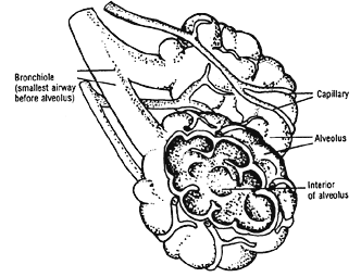
Figure 3. Each alveolar sac, or alveolus, is surrounded by one or more pulmonary capillaries. This alveolar-capillary unit is where oxygen (O2 ) and carbondioxide (CO2) are exchanged with the atmosphere. See Figure 4.
Figure 4. Cross-section of single alveolar-capillary unit. As blood flows past the alveolus, CO2 is given off and O2 is taken up. Each minute, under resting conditions, we breathe in about six liters of fresh air. About 1/3 of this air stays in the mouth, throat, and large airways where no gas exchange takes place; this region (the upper airways and part of our lungs) is referred to as "dead space," because air in this space doesn't take part in gas exchange. The remaining four liters of fresh air breathed in each minute are distributed to the hundreds of millions of alveoli and it this air that takes part in gas exchange and constitutes the alveolar ventilation. Pulmonary Circulation. Each minute our heart pumps approximately five liters of blood through the lung capillaries, distributing blood among the hundreds of millions of alveoli so gas exchange can take place. Because the lungs are three dimensional, one alveolar sac may be surrounded by several pulmonary capillaries. Each alveolus and all of its accompanying capillaries constitute the gas exchange unit (Figure 3). If there was no blood flow around the alveolus or there was blood flow without an accompanying alveolus, there would be no gas exchange. Thus alveolar ventilation is but one part of respiration; the other necessary part, the delivery of blood to the capillaries surrounding the alveoli, is the pulmonary circulation. The total circulation of the blood in our body is a circular affair. Blood flowing from one part of the heart to the lungs and back to another part of the heart constitutes one part of this circle; the other part is the systemic blood circulation, which is blood flowing from the heart to the rest of the body and then back to the heart (Figure 5). Arbitrarily, we can start this circle with one of the four chambers of the heart, the right atrium. Blood that enters the right atrium has given up much of its oxygen to the tissues, and is called venous blood (oxygen-poor or de-oxygenated blood). After collecting in the right atrium of the heart, venous blood then goes to the right ventricle from where it is pumped to the lungs in order to receive a fresh supply of oxygen. Blood leaving the right ventricle is pumped into the lungs via one large blood vessel, the main pulmonary artery. This large artery divides into two smaller pulmonary arteries, one to each of the lungs. Each pulmonary artery gives rise to many divisions, and in short order the blood supply pumped from the heart is divided among millions of tiny pulmonary capillaries, the smallest unit of circulation. These capillaries are in contact with the hundreds of millions of alveoli, the tiny sacs that receives the fresh air we inhale. The distance between each alveolus or air sac, and its surrounding capillaries, is very short, only the diameter of a thin membrane; oxygen easily diffuses from the air sac into capillary blood while, at the same time, CO2 diffuses out of the capillary blood and into the air sac. The CO2-laden stale air is then exhaled, and fresh air is brought into the lungs with the next breath. Oxygenated blood leaving the millions of pulmonary capillaries enters the other side of the heart, first the left atrium and then the left ventricle. From the left ventricle oxygen-rich blood is pumped, via the body's arterial circulation, to all the muscles, tissues and organs. In this way our kidneys, brain, liver, heart, bones, and other tissues receive vital oxygen. (When an analysis of arterial blood gas is performed for oxygen measurements, the blood sample is obtained by inserting a small needle into the radial artery of the wrist, just behind the thumb. Unlike venous blood, arterial blood reflects the status of gas exchange in the lungs and is therefore useful to examine in patients with many types of lung disease.)
Figure 5. Pulmonary and systemic blood circulation. RL=right lung; RV =right ventricle; RA=right atrium; VC=vena cava; PA=pulmonary arteries; LL=left lung; LV=left ventricle; LA=left atrium; PV=pulmonary veins; AO=aorta. The pulmonary and systemic (non-pulmonary) circulation systems are schematically shown in Figure 5. The heart, normally between our two lungs (Figure 1), is here separated to show its four chambers and the vessels leading to and from them. Each pulmonary artery branches into millions of tiny capillaries before picking up oxygen and giving off CO2 (gas exchange). Then the millions of capillaries merge to become the pulmonary veins. The pulmonary veins carry oxygenated blood back to the left heart, from where it is pumped, via the systemic arterial circulation, to the organs and tissues of the body. From these organs and tissues it then returns, via the systemic venous system, back to the right heart. In Figure 5 the light stipple represents venous or de-oxygenated blood and the dark stipple, arterial or oxygenated blood. Arrows show the direction of blood flow. Note that pulmonary arteries carry venous blood, and pulmonary veins carry oxygenated blood. The role of veins and arteries is reversed in the systemic circulation, where veins carry oxygen-poor (venous) blood and arteries carry oxygen-rich (arterial) blood. After entering the tissue, organ, or muscle, each systemic artery divides into smaller and smaller vessels, the smallest of which is the systemic capillary. These capillaries are structurally similar to the pulmonary capillaries and have the same function: to allow gas exchange to occur by simple diffusion. In the lung, oxygen diffuses into the capillaries and CO2 diffuses out. In all other capillaries (non-pulmonary or systemic capillaries) oxygen diffuses out of the capillary into the cells of the organ, and CO2 diffuses into the capillary from the cells of the organ. In this way oxygen is delivered for cellular metabolism and CO2, a waste product of metabolism, is removed. Gas exchange is a vital process; it occurs not only in the lungs but in all other tissues as well. Gas exchange requires both ventilation, provided by breathing adequate amounts of fresh air, and circulation, provided by the heart pumping blood to the lungs (pulmonary circulation) and then out to all other parts of the body (systemic circulation). Blood entering the systemic (non-pulmonary) capillaries is oxygenated. When blood leaves these capillaries it has given up some (not all) of its oxygen, and is venous or de-oxygenated blood. Venous blood, which appears blue in our veins under the skin, is actually dark red in a test tube. Arterial blood is normally bright red (although if the patient is low on oxygen arterial blood will also look dark red). The systemic capillaries, after delivering their oxygenated blood to the tissues, merge and form the veins that carry the venous blood; eventually all the systemic veins in the body come together to form the two great vena cavae, the superior, that carries blood leaving the head and neck, and the inferior, that carries blood leaving the rest of the body. Both vena cavae enter the heart at the level of the right atrium. Blood from the right atrium enters the right ventricle and then is pumped to the lungs to once again begin the process of oxygenation. The circle is completed.
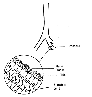
TEST YOUR UNDERSTANDING Answers DOES THE RESPIRATORY SYSTEM HAVE FUNCTIONS BESIDES GAS EXCHANGE? Yes. A particularly important respiratory system function is body defense. A healthy respiratory system prevents many airborne particles from damaging the alveoli or from entering the blood stream. The nose filters large airborne particles out of the air we inhale. This efficient filtering mechanism is bypassed, however, during mouth breathing. The trachea and main bronchi are also effective in keeping dusts and other large particles from reaching the alveoli. Coughing is one way we attempt to clear the large airways of noxious material. Even when our nose and normal cough response are not helping keep out dusts, our bronchi function silently to move out any unwanted material. This is accomplished by a blanket of mucus that covers the bronchi, and tiny hairs (cilia) which sweep the mucus out of the airways (Figure 6). When this dust-laden mucus reaches the top of the trachea, it is usually swallowed.
Figure 6. Each bronchus is lined with cells that are covered with tiny hairs, called cilia, that project into the airway. Cilia are covered with a blanket of mucus, which serves to collect dusts and other pollutants. This "mucus blanket," along with its collected dusts, is normally swept by the cilia up and out of the airways. Even if particles get past all of these defenses, special alveolar cells (called macrophages) are mobilized to help digest any foreign substances such as bacteria or tiny particles of dust. All of these normal defense mechanisms may be damaged from cigarette smoke, making smokers much more vulnerable to inhaled dusts and other impurities in the air. Answers to TEST YOUR UNDERSTANDING 1. b. oxygen in the atmosphere for carbon dioxide in the blood 2. oxygenated blood: a, c, d, g 3. leg vein --> inferior vena cava --> right atrium --> right ventricle --> lungs; normally these bubbles will be trapped by the lung capillaries. If any bubbles get through the lung capillaries they will take the path described in answer to the next question. 4. Pulmonary veins --> left atrium --> left ventricle --> aorta --> arterial circulation. REFERENCES AND BIBLIOGRAPHY Quoted sources and general references are listed by section or sections, in alphabetical order. An asterisk indicates references that are especially recommended. Medical textbooks and journal articles can be obtained from most public libraries via inter-library loan. For a list of companies that distribute free catalogs of diving books and videos, see Section U. See references for Section E. |


