 |
|
|
5. PetCO2 in lieu of PaCO2 in intubated patients
Summary: Measurement of end-tidal PCO2, called capnography, has been shown
to track PaCO2 in stable patients. Because the measurement requires a
closed system, PetCO2 monitoring works best in intubated patients.
Once a correlation is made between PetCO2 and PaCO2, the latter
need no longer be measured (or measured as frequently) in intubated patients,
including those being weaned from the ventilator.
|
 |
The figure shows a normal tracing of expired PCO2, measured during a tidal volume breath and printed out for placing in a patient's chart. Note the following about this figure:
In health, because carbon dioxide is never diffusion limited, alveolar PCO2 (PACO2) is assumed equal to the end-capillary PCO2 which in turn is equal to arterial PCO2 (PaCO2). Thus if you could sample alveolar PCO2 you would have a proxy for PaCO2. This in fact is possible because the last bit of air exhaled in each breath is the end-of-tidal-volume ('end-tidal') air and comes from the alveoli. By sampling exhaled gas breath-to-breath, we can obtain a continuous display of the end-tidal PCO2. This continuous reading should equal, or be very close (within 1-2 mm Hg) to the alveolar and arterial PCO2. To summarize:
In patients with lung disease and ventilation-perfusion (V-Q) imbalance, PetCO2 will likely not be equal or close to alveolar and arterial PCO2. This is because of increased physiologic dead space common in states of V-Q imbalance. Increases in dead space come about when a group of alveoli are un-perfused or under perfused, so that air reaching them becomes wholly or partly dead space air. Air in unperfused or under perfused alveoli keeps a low PCO2, since little or no CO2 is added to it (remember, inhaled air has almost zero CO2).
 |
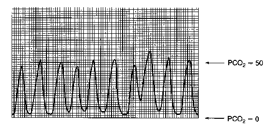
The above figure is a breath-by-breath tracing of exhaled CO2 from a patient with severe COPD. The patient was tachypneic so that the breaths appear compressed. Note the slight variation in PetCO2. In this patient the PetCO2 averaged approximately 50 mm Hg but his PaCO2 was 74 mm Hg, for a PaCO2-PetCO2 difference of 24 mm Hg. In this situation, the diseased alveoli do not empty evenly, and the end-tidal sample includes considerable dead space air.
There is no way to know how large the (PaCO2-PetCO2) will be in a given patient or a given type of lung disease. And while 24 mm Hg is a relatively high difference between PaCO2 and PetCO2, a large difference does not obviate usefulness of PetCO2. Like any blood gas measurement, the result must be validated and interpreted in light of the full clinical picture. The average PetCO2 in this sequence of breaths is about 50 mm Hg. The patient's PaCO2 measured at the same time was 74 mm Hg, for a PaCO2-PetCO2 of 24 mm Hg.
The principal advantages of capnography over intermittent measurement of arterial blood gases are: 1) to obviate the need for blood sampling; and 2) to allow for continuous reading of PetCO2. Thus, to the extent that PetCO2 can be used as a proxy for PaCO2, it has obvious utility in the intensive care setting. When properly used, PetCO2 (along with pulse oximetry) can often reduce arterial blood sampling and allow for safe patient monitoring.
There are two keys to properly using PetCO2: first, make a correlation with PaCO2 and second, know the pitfalls that can affect interpretation of PetCO2.
1) Correlate PetCO2 with PaCO2.
A sizable PaCO2-PetCO2 difference does not obviate the value of the PetCO2 for physiologic monitoring. A rise in PetCO2 still indicates a rise in PaCO2, but one cannot equate the numerical value of PetCO2 with PaCO2. Therefore, for physiologic monitoring at least one or two comparisons should be made between PetCO2 and PaCO2 before following PetCO2. Once the difference between the two values is established, and providing the patient remains clinically stable, the PetCO2 can be followed in lieu of the PaCO2.
Below are some data from a stable 38-year-old man receiving mechanical ventilation. Assuming there is no major change in his clinical condition, how could you use PetCO2 in weaning him from the ventilator?
|
Arterial blood gas and PetCO2 data FIO2 .40 pH 7.41 PaCO2 56 mm Hg PaO2 70 mm Hg SaO2 93% HCO3- 36 mEq/L
|
Note that his PetCO2 is 16 to 21 mm Hg lower than arterial PCO2. If he is stable, this difference can be added to the PetCO2 during weaning to determine PaCO2 at any time (ħa few mm Hg). If the patient remains clinically stable, one should not have to obtain an arterial sample to measure PaCO2. Of course other parameters should also be followed, such as pulse oximetry, respiratory rate, etc.
2) Know the pitfalls that can affect interpretation of PetCO2.
There are several potential pitfalls, but in the aggregate they are not common and should not dissuade one from using PetCO2 as a proxy for PaCO2 in intubated patients. While PetCO2 is not as widely used as is pulse oximetry, and is therefore a less familiar measurement, properly used it can be a valuable monitoring aid. Discussed below are three potential pitfalls.