Sarcoidosis: of Unknown Cause After 100 Years
WHAT IS SARCOIDOSIS?
Sarcoidosis is a systemic disease characterized by abnormal tissue swellings throughout the body. These swellings are called granulomas and are most commonly found in the lungs, liver, spleen, and skin. However, they may be found anywhere, including the eyes, ears, bones, and even in the coverings around the brain and spinal cord. Although these granulomas are usually widespread, they are very small and can only be seen with the aid of the microscope. When sarcoidosis appears on the skin it looks like raised rash or irritation; just by looking at the skin a physician can't make a definite diagnosis; the lesion has to be biopsied and examined under the microscope. The cause of sarcoidosis is completely unknown. In the past, organisms such as viruses and bacteria (similar to the tuberculosis bacteria) were suspected. At one time pine pollen was thought to be the cause since many of the cases were found in people who lived near pine forests. The evidence for these and other agents as a cure remains unproved. Sarcoidosis probably results from inhalation of some agent (?virus, ?bacteria), to which certain people react in an abnormal way by developing granulomas.
Sarcoidosis occurs in all ages and races and in both sexes. It does not favor any socioeconomic status, occupation, or level of education, and it is not caused by cigarette smoking, alcohol, or drugs. It is not inherited and does not tend to run in families. In the United States sarcoidosis seems to be most common among black women, but the reason is entirely unknown.
Many patients with sarcoidosis have no symptoms. The granulomas, hidden within the body's organs, are discovered only if carefully looked for. Thus, the clinical findings of sarcoidosis are nonspecific. In recent years certain radiologic and blood tests have proven more specific for this condition, so that some patients are now presumptively diagnosed without resort to tissue biopsy. However, for many patients a biopsy of some tissue is necessary to make the diagnosis.
The lungs are involved in over 90 percent of all
sarcoidosis cases, which is why pulmonary specialists are frequently
consulted. In fact sarcoidosis is usually first suspected by finding
an abnormal chest xray. It is unusual to see sarcoidosis
with no lung involvement, or where the chest xray is completely
normal. Historically, sarcoidosis was not always perceived as
a pulmonary condition. For many years after the disease was first
described in 1878, it was known as a skin condition only. In fact
this is the origin of its name; since the skin lesions so resembled
sarcomas (tumors) they were called "sarcomalike"
or "sarcoid." It was only in 1915, two decades after
the discovery of xrays by Roentgen in 1895, that sarcoidosis
was found to involve the lungs.
IS SARCOIDOSIS A SERIOUS ILLNESS?
So variable is sarcoidosis that generalization is difficult. Many people with sarcoidosis have no symptoms. The majority, even with symptoms, tend to suffer no severe impairment. Approximately 20 to 25 percent of patients do have permanent loss of lung function but are still able to live a relatively normal life. In only a small percentage of patients, less than five percent, is the sarcoidosis so severe as to eventually be fatal.
Symptoms are often referable to the respiratory system,
since the lungs are involved over 90 percent of the time. If pulmonary
sarcoidosis is extensive the patient may have shortness of breath,
but this is usually noticeable only on exertion, not at rest.
One of the most common reasons to treat sarcoidosis is for difficult
breathing. However, any time sarcoidosis causes impairment of
a vital organ, such as the eye or heart, treatment is also indicated.
HOW IS THE DIAGNOSIS OF SARCOIDOSIS MADE?
To be sure of the diagnosis in most patients, doctors must biopsy an involved organ or tissue and demonstrate sarcoidosis granulomas under the microscope. A major operation is almost never needed to obtain the biopsy. Some of the areas that may be involved with sarcoid granulomas, and from which tissue can be readily obtained, are the skin, the eyelid, the lungs, and the liver. Lung tissue may be biopsied by means of the fiber optic bronchoscope. In about 90% of pulmonary sarcoidosis cases, a tiny piece of lung tissue taken via the bronchoscope will suffice to make the diagnosis.
In many cases sarcoidosis can be diagnosed without resort to tissue biopsy. Generally this is accomplished when other likely diseases have been ruled out (such as tuberculosis) and the patient is not ill (patients with active tuberculosis or other infections are always ill with fever sweats, weight loss, fatigue, etc.). When patients with a chest x-ray typical of sarcoidosis are not ill or short of breath, we follow them without treatment. In the event of progression of disease (either on the chest xray or by development of symptoms), a tissue biopsy is generally needed.
Years ago a special skin test, known as the Kveim test, looked promising for the diagnosis of sarcoidosis. It involves taking a portion of the spleen from a patient with sarcoidosis and processing it in such a way that when injected under the skin of a suspected sarcoidosis patient a granuloma-type reaction occurs. Biopsy of the skin reaction area (after six weeks) and examination under the microscope reveals sarcoid granulomas. If the Kveim test was widely available and standardized it would be a helpful aid to make the diagnosis. Unfortunately, Kveim antigen (as the spleen extract is known) is available in only a few medical centers in the USA and is not standardized. In addition, the reaction itself is controversial since other diseases have been found to cause the same granuloma reaction. It seems the material injected has to be just the right chemical makeup or it won't be specific for sarcoidosis. Thus, the general unavailability of Kveim antigen and lack of standardization are reasons why it is rarely used.
Two noninvasive tools have proved helpful to make the diagnosis. One is the gallium scan, a nuclear medicine scan that may yield a highly specific pattern in patients with sarcoidosis. The patient is injected with Gallium-67 and scanned two days later, sarcoidosis tissues takes up the gallium and is highlighted in the scan. A typical pattern of Gallium uptake into the tissues may help diagnose sarcoidosis.
The other tool is a blood test called "Angiotensin
Converting Enzyme" or ACE. For unknown reasons the ACE level
is elevated in most active sarcoidosis cases. In fact an elevated
ACE level and characteristic gallium scan, along with a compatible
clinical history and chest xray picture, are often diagnostic
of sarcoidosis even without a tissue biopsy.
CAN SARCOIDOSIS MIMIC OTHER DISEASES?
Yes, sometimes. Among the diseases sometimes confused
with sarcoidosis are lymphoma (tumor of the lymph glands), tuberculosis,
and several unusual diseases that may cause the same xray
picture. When the diagnosis is uncertain and the patient is ill,
a tissue biopsy will be needed.
WHAT ARE THE STAGES OF SARCOIDOSIS?
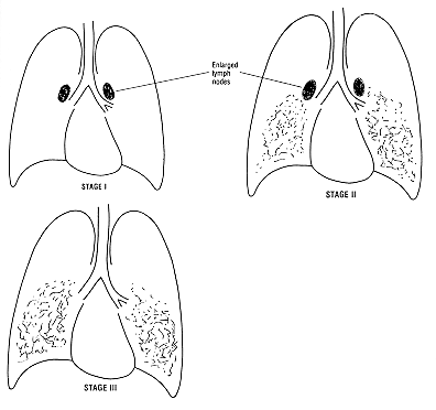
There are three classically described "stages" of sarcoidosis based on the chest xray: I, II, and III.
In Stage I the lymph nodes of the lungs near the center of the chest (the hilar areas) become enlarged, sometimes to the size of potatoes. The lungs themselves don't show any disease on the chest xray. Stage I patients usually have no symptoms and require no treatment (Figure 1a).
In Stage II there are enlarged lymph nodes, and in addition an abnormal pattern in the lung fields (Figure 1b). Stage II patients usually show some decrease in pulmonary function, as well as symptoms (cough or dyspnea); they may require treatment.
Stage III shows the lung infiltrates without evidence
of enlarged lymph nodes in the hilar areas (Figure 1c). It is
possible these patients once had the hilar "potato nodes,"
and that progression of their disease lead to lung involvement
with disappearance of the nodes. For most patients who present
with Stage III, however, this progression cannot be demonstrated.
Figure 1. Stages of Sarcoidosis

In actual practice, most patients don't show progression
from Stage I to III or even from Stage II to III. Patients may
present at any one of the three stages, and from that point may
stay there, improve (with disappearance of xray changes
altogether), or worsen (progress in staging). This is another
way of saying that the natural history of sarcoidosis is unpredictable.
WHAT IS THE TREATMENT FOR SARCOIDOSIS?
Most patients with sarcoidosis are not treated. Sarcoidosis tends to be a benign disease and its cause is unknown. The available treatment is not a cure, and is only helpful in slowing down (or occasionally stopping) the sarcoidosis process. Since this is nonspecific treatment for a disease of unknown cause, physicians only treat sarcoidosis patients if there is a compelling reason.
At present the only effective treatment for sarcoidosis is corticosteroids. The drug is usually taken by mouth as prednisone tablets. Steroids are also used in some forms of arthritis, for asthma and in many other medical illnesses. For sarcoidosis, steroids are considered nonspecific treatment. Limited evidence suggests that steroid treatment, once begun, has to be given for at least several months to be effective. The aim of treatment is to improve the patient's function or to at least slow down progression of the sarcoidosis. The dose of prednisone, and how long to treat, are controversial. Some physicians recommend high doses (40 mg a day or more) for long periods, others low doses (15 mg or less) for shorter periods.
Unfortunately, sarcoidosis can never be considered cured, only slowed down or arrested. There are several reasons for this statement. First, in its early stages the disease sometimes gets better without any treatment whatsoever. Thus, physicians are never sure if a treated patient improved because of the steroids or because of a natural remission. Second, some patients improve after taking the steroid medication, but when they stop the drug, suffer a relapse — the sarcoidosis flares up. Third, even after prolonged steroid treatment microscopic sarcoid granulomas can often be found in various organs, indicating that some stage of the disease is still present.
Progressive sarcoidosis, unresponsive to steroids,
is a medical dilemma. There is currently no other good treatment
for the disease, although certain "auto-immune" drugs
are sometimes used such as Cytoxan or Imuran. Any patient who
has progressive or severe sarcoidosis unresponsive to steroids
is likely to have complications, either of the disease or of treatment,
and should come under the care of a physician experienced in managing
the problem. An example is the patient with advanced sarcoidosis
who has developed respiratory failure, or the patient who has
metabolic derangement from long-term, high dose steroids. Fortunately,
such cases represent a small minority of all sarcoidosis patients.
Mrs. B. – A Case of Sarcoidosis
not requiring Treatment
Mrs. B. is a 24-year-old elementary
school teacher who had a routine chest x-ray as part of her pre-employment
physical. The chest x-ray revealed large "potato nodes"
in both lungs. She was referred to a physician for evaluation
who learned that Mrs. B. had no symptoms whatsoever; no fever,
weight loss or other medical problems. She felt healthy and well.
She never had a chest x-ray before. Skin tests for tuberculosis
were negative. Gallium scan showed a typical uptake pattern consistent
with sarcoidosis. Because she had no symptoms and had a typical
chest x-ray and Gallium scan, the diagnosis of sarcoidosis was
presumed. A letter was written to the school board stating that
she had sarcoidosis and that it was not contagious, not infectious,
and that it should not affect her work. However, because of concern
about disease progression, she was followed with serial chest
x-rays every 6 months. Within a year her chest x-ray showed complete
resolution of the nodes. Mrs. B had a case off spontaneous regression
of Stage I Sarcoidosis.
T.R.S. – A Case of Sarcoidosis
Requiring Treatment
Mr. S. is a 27yearold graduate student who has had a persistent dry cough for the past month. He has also lost about 10 pounds. A chest xray in the college health clinic shows a diffuse lung infiltrate and enlarged hilar lymph nodes in both lungs (Stage II Pulmonary Sarcoidosis). He is referred to a chest physician.
On further history we learn he has not been exposed to toxins, chemicals, or tuberculosis (TB). He has no personal history of TB or other lung disease and has never smoked. His last chest xray, four years ago, was normal.
He is admitted to the hospital for further tests. Skin tests show no reaction to TB or mumps; since most of the population reacts to a mumps skin test, this result suggests a depression in his immune system, a common finding in sarcoidosis. Blood tests are normal, helping to rule out sarcoid involvement of his kidneys, liver, or pancreas. Pulmonary function tests reveal some decrease in his vital capacity, consistent with sarcoidosis. A 48-hour gallium scan shows increased activity in both lungs, consistent with active sarcoidosis. All of these studies are completed within 48 hours and a presumptive diagnosis of sarcoidosis is made. Because he has symptoms (cough and weight loss) and warrants treatment, an attempt is made to obtain diagnostic tissue. On the third hospital day fiber optic bronchoscopy and trans-bronchial lung biopsy are performed. The results show noncaseating granulomas, confirming the diagnosis of sarcoidosis .
He is started on oral prednisone and
within three days his cough disappears and he feels better. He
is prescribed low dose prednisone for the next 12 months and plans
are made to follow his course with serial chest xrays and
breathing studies .
Mrs. Z. – A Case of Progressive
Sarcoidosis
Mrs. Z. is a 54-year-old woman who first
had sarcoidosis diagnosed 12 years earlier. Since then her chest
x-rays have showed progression of the disease and she has become
increasingly short of breath. She was started on prednisone 8
years earlier and took the drug for 3 years, but then quit it
because of severe side effects, including some weakening of her
bones and diabetes. Because the disease continued to progress
however, steroids were re-started, albeit at a lower dose, two
years earlier. She has been on low-dose Prednisone for two years.
She is now very limited and cannot walk without the aid of oxygen.
Lately her cough has recurred and she complains of having non-specific
chest pains. Pulmonary function studies show very severe impairment,
and she qualifies for Social Security disability. Thorough review
of her records reveals no history of tuberculosis or any other
disease that could possibly have caused her current condition.
She has end-stage lung disease, attributable to progressive sarcoidosis.
[Return to Table of Contents]